Introducción
Epidemiología y generalidades
La incidencia de estas fracturas es de un 2%, teniendo en cuenta que las fracturas localizadas en el codo suponen un 7% y, dentro de las fracturas de húmero, las que afectan a su porción distal suponen aproximadamente un 30% de todas las fracturas(1) En cuanto a la edad y el sexo, tienen una distribución bimodal, en jóvenes son más frecuentes en varones, mientras que en ancianos se asocian más a mujeres con osteoporosis.
Mecanismo lesional
• En jóvenes se producen por accidentes de alta energía (accidentes laborales, deportivos, de tráfico, etc.). Suelen ser fracturas más complejas, supraintercondíleas conminutas.
• En ancianos, se suelen producir por accidentes de baja energía, como caídas desde su propia altura o traumatismos indirectos con hueso osteoporótico.
• En niños, suelen darse por caídas sobre la mano con el brazo en extensión. Son más frecuentes las supracondíleas extraarticulares.
Anatomía
El húmero distal se proyecta distalmente hacia anterior, en un ángulo de 45°; además, tiene un valgo fisiológico de unos 6°. Del mismo modo, el cúbito proximal también presenta dicha angulación, lo que va a permitir que, incluso en flexión máxima, haya un espacio entre ambos huesos, facilitando así un lugar para que se alojen las masas musculares. Esta angulación también permite que la apófisis coronoides no choque con la fosa coronoidea hasta que ambos huesos no estén paralelos(2).
Además, existe un cambio en la morfología del húmero distal, con una forma más cilíndrica en su porción proximal, mientras que se aplana en su porción distal, formando las crestas supracondíleas interna y externa que finalizan en los cóndilos interno y externo, y su porción más prominente, los epicóndilos. La columna medial del húmero distal es más pequeña que la lateral y más prominente, siendo más frágil y estando más predispuesta a fracturas frente a traumatismos o procedimientos quirúrgicos(1).
La superficie articular del húmero distal está formada por:
• La tróclea humeral: con forma de polea, presenta 2 carillas convexas que van a permitir su articulación con el cúbito proximal, que tiene forma de “silla de montar”.
• El cóndilo humeral o capitellum: es una hemiesfera anterior sin continuidad posterior, cuya curva de convexidad es la misma que la curva de concavidad que presenta la cabeza del radio, formando así la articulación húmero-radial. Esto va a permitir que, sea cual sea el grado de flexoextensión del codo, la cúpula radial pueda pivotar sobre el cúbito, permitiendo así la pronosupinación.
El codo presenta una serie de estabilizadores(3) para los distintos arcos de movimiento:
• Cápsula articular: se extiende a lo largo de toda la articulación del codo, anterior y posteriormente, actuando como un estabilizador estático.
• Ligamentos: el ligamento colateral medial (cubital) es el principal estabilizador del codo en el valgo, mientras que el complejo ligamentoso lateral (sobre todo el ligamento colateral lateral cubital) actúa como el estabilizador fundamental en el varo.
• Músculos: la musculatura flexora, flexopronadora y extensora del codo actúan como estabilizadores dinámicos, favoreciendo la congruencia articular y dando soporte en todos los arcos de movimiento.
La arteria humeral desciende por el brazo hasta situarse medial al músculo braquial, pasando posteriormente medial al tendón del bíceps y cruzando la articulación del codo, dando una abundante circulación colateral, para después dividirse en las arterias radial y cubital.
Además, todos los nervios periféricos del miembro superior cruzan la articulación del codo:
• El nervio mediano recorre el brazo medial a la arteria humeral y cruza el codo entre las 2 cabezas del pronador redondo, hasta situarse entre el flexor superficial y el flexor profundo de los dedos, emitiendo posteriormente el nervio interóseo anterior (NIA). A lo largo de su recorrido, inerva a la musculatura flexora del compartimento flexor superficial y puede verse frecuentemente lesionado en las fracturas supracondíleas de codo.
• El nervio cubital discurre posterior a la epitróclea humeral y cruza el codo entre las 2 cabezas del flexor cubital del carpo.
• El nervio radial atraviesa la articulación del codo, en posición anterior con respecto al epicóndilo, entre los vientres musculares del braquial y el braquiorradial, para posteriormente dar su rama sensitiva (el nervio radial propio) y motora (nervio interóseo posterior). Este último tiene relevancia en los abordajes anteriores y externos, pues tiene riesgo de lesionarse a nivel del radio proximal, por lo que será importante supinar el antebrazo para desplazarlo hacia el lado externo en el primer caso y situar el antebrazo en pronación en el segundo caso.
Clasificación
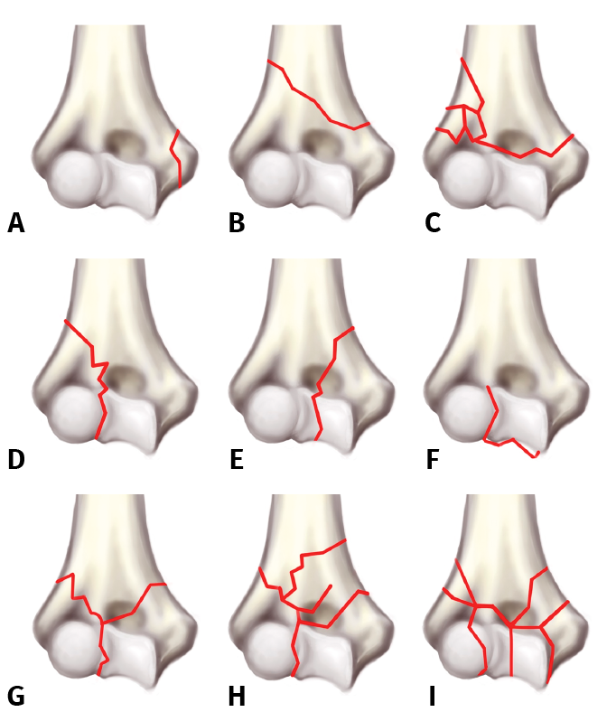
Existen varias clasificaciones, la clasificación de la AO Foundation/Orthopaedic Trauma Association (AO/OTA) es una de las más usadas. En dicha clasificación, cualquier fractura del húmero se nombra con el número 1 y al localizarse en su porción distal se sigue con el número 3. Por lo tanto, todas comenzaran con la nomenclatura 13 (húmero 1, distal 3) y se subclasifican en A, B o C según el tipo de fractura:
A. Extraarticular:
A1. Avulsión apofisaria (Figura 1A).
A2. Fractura metafisaria simple (Figura 1B).
A3. Fractura metafisaria multifragmentaria (Figura 1C).
B. Articular parcial:
B1. Sagital externa (Figura 1D).
B2. Sagital interna (Figura 1E).
B3. Frontal (coronal): (Figura 1F).
C. Articular completa:
C1. Articular y metafisaria simple (Figura 1G).
C2. Articular simple y metafisaria compleja (Figura 1H).
C3. Articular y metafisaria compleja (Figura 1I).
Figura 1. A: extraarticular. Avulsión apofisaria; B: extraarticular. Fractura metafisaria simple; C: extraarticular. Fractura metafisaria multifragmentaria; D: articular parcial. Sagital externa; E: articular parcial. Sagital interna; F: articular parcial. Sagital interna; G: articular completa. Articular y metafisaria simple; H: articular completa. Articular simple y metafisaria compleja; I: Articular completa. Articular y metafisaria compleja.
A su vez, se pueden clasificar siguiendo una denominación anatómica, como en la clasificación de Muller, que se basa en la afectación articular(4):
• Extraarticulares: suponen el 10% de las fracturas de húmero distal, son el tipo A de la clasificación de la AO y, según su localización, se pueden denominar:
– Supracondíleas: el trazo de la fractura es proximal a los cóndilos y a la articulación.
– Diacondíleas o transcondíleas: el trazo es paralelo a la anterior pero más distal, afectando a los cóndilos (cóndilo y tróclea humeral). Son más frecuentes en ancianos.
• Parcialmente articulares: suponen un 5%. Son el tipo B de la clasificación de la AO. Según afecten a la columna medial o lateral, se denominan:
– Condilares:
- Tipo I: es una avulsión, no provoca inestabilidad.
- Tipo II: afecta a todo el cóndilo, puede provocar inestabilidad.
– Trocleares:
- Tipo I: es una avulsión, no provoca inestabilidad.
- Tipo II: afectan a la tróclea. Pueden provocar inestabilidad.
• Articulares: se denominan supraintercondíleas, suponen un 60% y tienen trazo extraarticular y articular, es decir, no hay continuidad entre los fragmentos articulares y la diáfisis. Son el tipo C de la clasificación de la AO.
Dependiendo del grado de desplazamiento, rotación y conminución de sus fragmentos, a su vez, se clasifican, según Riseborough y Radin(5), en:
– Tipo I: no desplazada.
– Tipo II: fragmentos desplazados, pero no rotados.
– Tipo III: fragmentos desplazados y rotados.
– Tipo IV: conminución y gran desplazamiento de los fragmentos.
• Fracturas del capitellum: son fracturas poco frecuentes que solo afectan a la porción articular del cóndilo, sin verse afectadas las inserciones ligamentosas ni las inserciones musculares.
Su clasificación es la de Bryan-Morrey:
– Tipo I: Hahn-Steinhal, afecta a gran parte del cóndilo, incluyendo cartílago y porción ósea.
– Tipo II: Kocher- Lorenz, afecta solo al cartílago condilar, es una fractura subcondral.
– Tipo III: es una fractura del cóndilo conminuta.
– Tipo IV: se extiende a la tróclea.
• En niños, la clasificación más usada es la de Gartland, que clasifica fracturas supracondíleas extraarticulares con el fragmento distal en flexión (2%) o en extensión (98%). Según el grado de desplazamiento del mismo:
– Tipo I: no desplazada.
– Tipo II: ligeramente desplazada con una cortical íntegra, ligeramente rotada/angulada.
– Tipo III: desplazamiento completo de la extremidad distal, posteromedial, posterolateral, anterolateral.
Diagnóstico
Exploración clínica
El paciente suele presentar dolor asociado a impotencia funcional. En el examen físico se deben palpar las prominencias óseas: olécranon y epicóndilos lateral y medial, que en condiciones normales forman un triángulo equilátero (triángulo de Nelaton). Es importante prestar atención al estado de las partes blandas, ya que condicionarán el tratamiento y la exploración neurovascular, que deberán ser evaluadas con cuidado y convenientemente documentadas.
Habitualmente, existe tumefacción, hematomas o arrugas de la piel, debidas estas últimas a que el segmento proximal puede perforar el músculo braquial anterior y comprimir la dermis profunda (pucker sign). Ante este signo, existe una alta probabilidad de complicaciones neurovasculares por lesión de la arteria humeral, nerviosa o presencia de síndrome compartimental.
En algunos casos coexiste la presencia de inestabilidad, que puede condicionar la presencia de lesiones neurovasculares. Hasta un 49% de los pacientes suelen presentar complicaciones neurovasculares(6), por lo que es importante hacer un examen detallado de las funciones motoras y sensitivas de los nervios mediano, radial, cubital, interóseo anterior e interóseo posterior. Además, hay que evaluar la perfusión del miembro superior, puesto que en las fracturas más complejas puede lesionarse la arteria humeral (hasta el 38% de los casos según Campbell et al.)(7). De esta forma, el estado vascular puede clasificarse en 3 grados en función de la presencia o ausencia de pulso y perfusión: clase I o buena perfusión distal (mano caliente y roja con pulso radial presente); clase II (buena perfusión distal, pero con ausencia de pulso radial); y clase III (mano fría, azul o pálida y con ausencia de pulso radial). Tanto las lesiones de clase II como las lesiones de clase III requieren un manejo urgente.
Pruebas de imagen
El estudio radiológico comenzará por unas radiografías simples de codo, tanto la proyección anteroposterior (AP, tomada con el codo flexionado a unos 40°) como la proyección lateral nos darán una idea del tipo de fractura, la conminución y el grado de desplazamiento en los distintos ejes del espacio.
Existen algunos signos característicos en la radiología simple que, si se conocen, pueden ser útiles para sospechar algunas lesiones y profundizar en el estudio radiológico evitando que pasen desapercibidas. Algunos ejemplos de ellos son: el signo de la vela(8), que se corresponde con el aumento de volumen en la almohadilla grasa anterior o posterior de la articulación del codo, en relación con la presencia de hemartros o derrame sinovial. Este signo puede ser útil para sospechar fracturas no evidentes radiológicamente. Cabe distinguir que la presencia de líquido en la almohadilla grasa anterior es muy sensible pero poco específica, puesto que cualquier lesión que provoque un desplazamiento de esta almohadilla (por ejemplo, un derrame sinovial) puede hacer que sea visible en la radiografía, ya que es mucho menos profunda que la almohadilla posterior. Sin embargo, la presencia de derrame en la almohadilla grasa posterior es más específica de una fractura intraarticular, puesto que necesita un mayor volumen para verse desplazada de la fosa olecraneana.
También la presencia del signo del “arco doble” en una radiografía lateral pura del codo es patognomónico de una fractura del capitellum con trazo coronal que suele extenderse a la tróclea.
La tomografía axial computarizada (TAC) es imprescindible en el estudio, ya que nos va a permitir definir el patrón óseo de fractura, la afectación articular y permite una mejor planificación preoperatoria.
La resonancia magnética nuclear (RMN), sin embargo, no suele emplearse para el diagnóstico en las lesiones agudas.
En caso de sospechar lesiones nerviosas, habría que solicitar un electromiograma y, en caso de lesiones vasculares, una angio-TAC o arteriografía urgente, además de consultar con cirugía vascular.
Tratamiento
Opciones de tratamiento. Indicaciones y contraindicaciones
El tratamiento quirúrgico será el tratamiento de elección para la gran mayoría de las fracturas de húmero distal. Los objetivos del tratamiento son restaurar la anatomía del húmero distal, la superficie articular y aportar una fijación estable que permita la movilización precoz y evite rigideces. Restaurar la función del codo es el propósito principal, aunque en muchos de estos casos resulta técnicamente difícil por la complejidad de la lesión.
El tratamiento de este tipo de fracturas ha ido evolucionando mucho en los últimos años. Gracias al uso más extendido de la TAC, es posible hacer una reconstrucción tridimensional que aporte más información sobre el patrón de fractura, diagnosticar de forma más precisa la afectación articular y realizar la planificación quirúrgica. También avances con respecto al material y la técnica, como entender los beneficios del sistema de placas paralelas entre sí, el uso de placas periarticulares preconformadas y el aumento en cuanto a forma, tamaño y tipo de placas disponibles que permiten una mejor adaptación a la anatomía. Otra reciente aportación de la tecnología ha sido la impresión 3D, la cual, con imágenes de TAC del codo fracturado y del codo contralateral sano, permite hacer un modelo tridimensional del húmero distal sobre el que se puede diseñar la reconstrucción, planificar la colocación de las placas, elegir el tipo de placa y premoldearlas con antelación.
El tratamiento conservador se reserva para pacientes mayores con baja demanda funcional y contraindicaciones absolutas para la anestesia o la cirugía.
La artroplastia de codo tiene indicación en casos seleccionados de pacientes con edad avanzada con baja demanda funcional y patología articular preexistente, tal como artritis reumatoide, artrosis, etc. También, en casos de fracturas muy complejas con extensa conminución y daño articular severo que tengan osteopenia importante. También se utiliza en casos de salvamento de osteosíntesis fallidas(9,10). Es excepcional la utilización de la artroplastia en el entorno de la traumatología laboral, ya que limita la realización de tareas de fuerza y, por ello, suelen indicarse en pacientes con baja demanda funcional.
Tratamiento quirúrgico
Vías de abordaje
Existen distintas vías de abordaje dependiendo de las características de la fractura que se va a abordar y el tipo de tratamiento:
• El abordaje posterior es el más frecuente, consiste en realizar una incisión sobre la línea media, ligeramente medial a la punta del olécranon. A través de esta incisión se accede bien al nervio cubital, para liberarlo y protegerlo con un vessel-loop durante la cirugía. La transposición subcutánea anterior del nervio cubital es recomendable, ya que reduce el contacto del nervio sobre la placa medial y permite proteger el nervio durante la cirugía, pero hay cirujanos que prefieren no transponerlo, lo cual también es una opción válida siempre y cuando no contacte con el material de osteosíntesis. Se puede realizar con o sin osteotomía del olécranon, como se desarrolla a continuación.
La osteotomía del olécranon suele hacerse para obtener una visión óptima de la superficie articular. La más frecuentemente utilizada es la de tipo chevron, con 2 cortes oblicuos sobre el olécranon que convergen en el vértice distal con forma triangular. Se inicia con una sierra y se completa con un escoplo para evitar dañar con la sierra el cartílago del húmero que está adyacente y minimizar la pérdida de hueso. Existen descritas otras técnicas de osteotomía del olécranon –Muller, MacAusland–, en las que varía la forma de la osteotomía pero el concepto es el mismo.
En casos en los que la fractura sea más simple y no tenga afectación articular, puede evitarse la osteotomía, realizando otros abordajes como el paratricipital bilateral (Alonso Llames), transtricipital o desinsertando el tríceps del olécranon en el Bryan- Morrey. Estos son los abordajes posteriores más utilizados, aunque existen más descritos.
• Abordajes laterales: los más frecuentes son el abordaje de Kaplan y el Kocher, que se utilizan entre otras indicaciones para fracturas condilares aisladas del cóndilo lateral. El abordaje de Kaplan va en el intervalo entre el extensor radial del carpo largo (ERCL) y el extensor común de los dedos (ECD), y el Kocher va entre el ancóneo y el extensor ulnar del carpo (ECU); este último tiene menos riesgo de lesión del nervio interóseo posterior.
• Y los abordajes mediales se utilizan para fracturas del cóndilo medial. El más frecuentemente utilizado es el abordaje de Hotchkiss, en el intervalo entre el flexor cubital del carpo y el pronador redondo.
Técnicas quirúrgicas
Osteosíntesis con placas
El triángulo de estabilidad en el húmero distal está formado por las columnas medial, lateral y la masa transcondílea. El objetivo es restaurar esas 3 columnas(11); para ello, la técnica quirúrgica más utilizada y que ha demostrado ser la que más estabilidad aporta es la osteosíntesis con 2 placas(12,13). El uso de una sola placa reduce la estabilidad del montaje. Otras técnicas como agujas o tornillos aislados están contraindicados por falta de estabilidad, excepto en fracturas condilares aisladas.
Dentro de las osteosíntesis con 2 placas, existe debate entre colocarlas paralelas entre sí o la configuración clásica ortogonal (90-90) colocando una medial y otra en la cara posterior de la columna lateral. En los últimos años se ha demostrado que la configuración de placas paralelas, en la que se colocan tornillos bloqueados de ángulo fijo, mediales y laterales interdigitados entre sí, de forma que todos agarran fragmentos del lado opuesto, es el montaje biomecánicamente más estable, ya que asemeja a un arco(13,14).
Manejo de la pérdida ósea
En casos complejos de fracturas con mucha conminución será necesario resecar pequeños fragmentos óseos sin vascularización imposibles de reconstruir, lo que causará pérdida ósea.
Si esta pérdida ósea se produce a nivel supracondilar, puede tratarse asumiendo un acortamiento del húmero, que hasta 2 cm tiene mínimo impacto en la biomecánica del codo(15). El borde de la diáfisis distal se puede contornear para que afronte regularmente con el fragmento de húmero distal y poder hacer compresión. El riesgo de realizar esta técnica es el malalineamiento rotacional y en varo-valgo, que debe comprobarse antes de la fijación definitiva. Además, pérdidas de espacio en las fosas olecraneana y coronoidea pueden producir pérdidas del rango de movilidad en flexión y en extensión; para evitarlo, se debe angular ligeramente hacia anterior el fragmento distal, para crear la fosita coronoidea, y se puede crear la fosita olecraneana con ayuda de una fresa redonda u ovalada de alta velocidad.
Si la conminución se localiza en el centro del húmero distal, se puede colocar injerto óseo. El cartílago del aspecto central de la tróclea no es esencial, el del capitellum y el aspecto medial de la tróclea sí debe ser preservado. El injerto óseo puede ser autoinjerto de cresta ilíaca, aloinjerto parcial osteoarticular de húmero distal.
Osteosíntesis con tornillos
Las fracturas aisladas que afectan al cóndilo o a la tróclea se pueden tratar mediante abordaje lateral o medial y tornillos de compresión sin cabeza(16).
En casos de afectación articular, con fragmentos pequeños no sintetizables, se puede extirpar el fragmento e iniciar una movilización precoz. Si la afectación articular es más extensa, se tendrá que valorar la artroplastia.
Fijación externa
El fijador externo permite la reducción por ligamentotaxis; es un método de fijación provisional indicado en casos con mucha afectación de partes blandas o fracturas abiertas que requieran técnicas de cobertura, hasta que las partes blandas permitan realizar una fijación definitiva. También en casos que asocien inestabilidad.
Manejo posquirúrgico
En el postoperatorio inmediato se coloca una férula posterior a 90°, para reducir el dolor y el edema de las partes blandas, pero en cuanto sea posible hay que iniciar la movilización, puesto que el codo es una articulación con tendencia a la rigidez. Si la síntesis es estable, en la mayoría de las fracturas intervenidas con doble placa se puede retirar la férula a los 10-15 días de la cirugía para introducir ejercicios de flexoextensión activa autoasistida con el brazo contralateral sano, tan pronto como sea posible.
La presencia de lesiones ligamentosas asociadas a las fracturas demora la introducción de la movilidad completa hasta las 6 semanas, por riesgo de inestabilidad recurrente. En casos con lesiones ligamentosas se recomienda utilizar ortesis de movilización activa, las cuales tienen una rótula en la que se pueden bloquear los movimientos que se quieran proteger en función de qué ligamento esté afectado. De esta manera, se limitan algunos grados del rango articular de los movimientos que se quieran evitar y se van aumentando progresivamente.
La carga de pesos y el fortalecimiento deben demorarse hasta que la fractura esté consolidada, que será como mínimo pasadas 6-8 semanas, pero debe confirmarse la consolidación radiológica de la fractura antes de su introducción.
Tratamiento conservador
El tratamiento conservador está raramente indicado en este tipo de fracturas. Se reserva para pacientes con mucha patología de base que suponga una contraindicación absoluta de la cirugía o de la anestesia.
También en casos de osteoporosis severa en los que sea esperable un aflojamiento y fracaso de la osteosíntesis.
Otra indicación serían casos de fracturas en las que coexista mucha afectación o destrucción de las partes blandas (explosiones, aplastamientos…); en estos casos se plantea la opción conservadora.
El tratamiento conservador consiste en inmovilización con férula de yeso o yeso braquial hasta que radiológicamente se objetive consolidación de la fractura o con suficiente estabilidad como para comenzar la movilidad sin riesgo de desplazamientos.
En casos de ancianos con hueso muy osteoporótico, conminución y que vayan a tolerar mal el yeso, se plantea el cabestrillo en “saco de huesos”, con el codo en máxima flexión para que se alinee la fractura por ligamentotaxis.
Debido al tiempo de inmovilización, cabe esperar que se produzca rigidez articular del codo, la cual es preferible a otras complicaciones como la pseudoartrosis dolorosa o infectada, complicaciones temibles del tratamiento quirúrgico.
Complicaciones
La neuropatía cubital es una de las complicaciones más frecuentes; se produce aproximadamente en un 15% de las fracturas intervenidas mediante osteosíntesis. En la mayoría de los casos son neuroapraxias transitorias que se resuelven con el tiempo, pero es recomendable realizar electromiografía de control(17).
Molestias derivadas del material, en pacientes con poco tejido subcutáneo y zonas de prominencia ósea, como el olécranon, el epicóndilo y la epitróclea, suelen resolverse con una retirada del material cuando haya consolidado la fractura, habiendo pasado entre 6 y 12 meses dependiendo del tipo de fractura.
La rigidez es bastante frecuente en la fase inicial, aunque la síntesis haya quedado estable y se introduzca de forma temprana la movilidad. La inflamación y el dolor postoperatorios limitan al paciente para realizar ejercicios de movilidad y en la mayoría de los casos esa demora más la fibrosis producida por la cicatrización de las partes blandas suelen limitar el rango articular. Lo esperable en la mayoría de los casos es que sea transitoria y se resuelva con rehabilitación; en los casos en los que no sea así, puede plantearse una artrolisis, asociada o no a la retirada del material, siempre y cuando la fractura haya consolidado suficientemente.
Pseudoartrosis: en caso de no conseguir consolidación tras haber esperado un tiempo prudencial, estaría indicada una nueva osteosíntesis con injerto óseo y placas de compresión.
La malalineación o malrotación pueden corregirse con osteotomías.
Conclusiones
Las fracturas de húmero distal suponen un tercio de todas las fracturas que afectan al húmero (30%). Su mecanismo lesional varía en función de la edad del paciente, así como el patrón de fractura más frecuente. También en la indicación del tratamiento, es un factor importante la edad, así como la demanda funcional del paciente, el tipo de fractura, la calidad ósea y el estado de las partes blandas.
En general, el tratamiento de las fracturas de húmero distal es eminentemente quirúrgico. Su principal objetivo es restaurar la anatomía y aportar estabilidad para permitir una movilización precoz. Pero cabe tener en cuenta que son fracturas técnicamente difíciles y con complicaciones asociadas.
Desde el punto de vista laboral, hay que esperar que son lesiones graves, con secuelas y tiempos de recuperación prolongados.